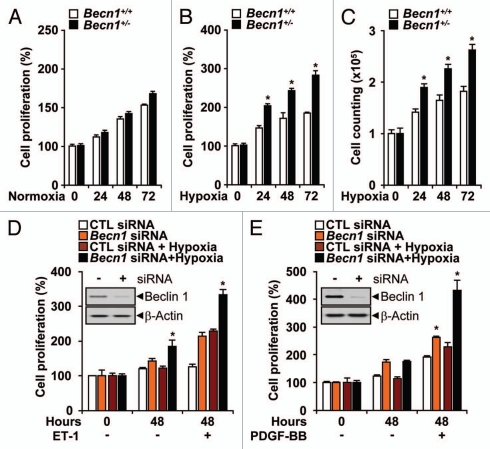
Figure 4.
Heterozygous disruption of Becn1 promotes hypoxia-induced endothelial cell proliferation, migration and tube formation. (A) The proliferation of Becn1+/+ and Becn1+/− mLECs exposed to normoxia was assessed by crystal violet staining. (B and C) Proliferation indices of mLECs exposed to hypoxia (1% O2) were assessed by crystal violet and Trypan blue exclusion methods. Data represent means ± SEM from experiments in triplicate (*p < 0.05, Student's t-test). (D and E) PAECs or PASMC were transfected with control or Becn1 siRNA. Western (inserts) shows validation of Beclin 1 expression levels following control or Becn1 siRNA treatment. β-actin served as the standard. (D) Following addition of ET-1 (40 nM), PAEC cultures were exposed to hypoxia or normoxia for an additional 48 h. Data represent means ± SEM in triplicate experiments. (*p < 0.05, Student's t-test). (E) Following addition of PDGF-BB (20 ngml−1) siRNA transfected PASMC cultures were exposed to hypoxia or normoxia for an additional 48 h. Data represent means ± SEM from three independent experiments. (*p < 0.05, Student's t-test).

