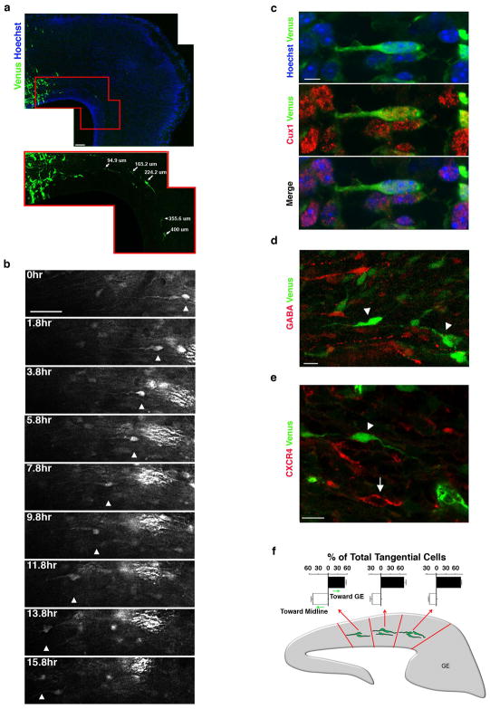
Figure 3. Lpd depleted bipolar pyramidal neurons migrate tangentially within the IZ/SVZ but do not exhibit a change in cell fate.
(a) E18.5 cortical sections electroporated at E14.5 with Lpd shRNA and Venus. Cells were counterstained with Hoechst 33342 dye to visualize nuclei (blue). Enlargement of boxed region is shown below. The distance between the cell body of individual tangential cells and the border of the electroporated region is indicated (arrows). (b) Time-lapse of a tangentially oriented bipolar cell migrating in the IZ/SVZ (arrowhead). (c) Image of a tangentially oriented bipolar cell, co-electroporated with Lpd shRNA and Venus, that expressed the neuronal marker, Cux1 (red). Nuclei were visualized with Hoechst 33342 dye. Cortical neurons expressing Lpd shRNA and Venus (arrowheads) lack detectable (d) GABA expression (red), and (e) CXCR4 (red) expressed by migrating interneurons (arrow). (f) Orientation of the leading process of Lpd-depleted tangentially oriented bipolar cells in the medial, mediolateral, and lateral regions of the cortex. Brains were electroporated at E11.5 and harvested 37 hrs later. Bar graphs are plotted as mean ± SEM. Scale bars: 50 μm (a), 40 μm (b) 5 μm (c), and 10 μm (d) and 10μm (e).

