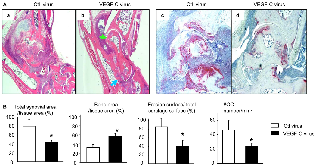
Figure 4. AAV-VEGF-C reduces joint tissue damage in TNF-Tg mice.
Four months after virus injection, legs were harvested and subjected to histologic analysis. (A) Representative H&E (×2) and TRAP (×4) stained sections show decreased joint tissue damage, including cartilage erosion (green arrows) and bone loss (blue arrows), and TRAP+ osteoclasts in the VEGF-C-treated joints. (B) Quantitation of synovial volume, bone area, cartilage erosion, and osteoclast numbers. Values are the mean ± SD of 5–6 legs per group. *p<0.05 vs control virus.

