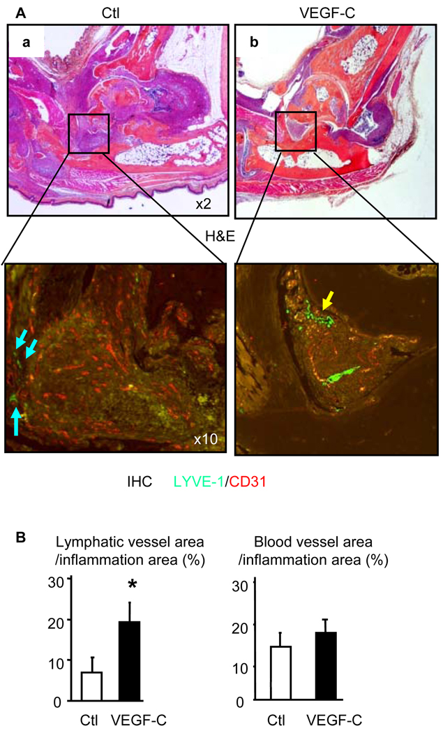
Figure 6. AAV-VEGF-C increases lymphangiogenesis in joints of TNF-Tg mice.
(A) Four months after AAV injection, legs were harvested and subjected to double immuno-fluorescence staining with anti-LYVE-1 and anti-CD31 antibodies. Representative H&E (×2), LYVE-1 (green-lymphatic vessels, ×10) and CD31 (orange-blood vessels, ×10) stained ankle sections show that LYVE-1+ lymphatic vessels are present around synovium (blue arrows) in control virus treated legs or within the synovium (yellow arrows) in VEGF-C-treated legs. (B) Quantitation of LYVE-1+ lymphatic and CD31+ blood vessel area inside the areas of inflammation. Values are the means ± SD of 5–6 legs per group. *p<0.05 vs. control virus.

