
Figure 2. Chronic intermittent ethanol exposure and withdrawal upregulate the synaptic function of AMPA-type glutamate receptors in the lateral/basolateral amygdala.
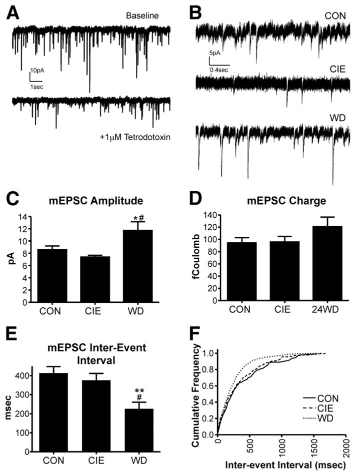
The figure is republished with permission of the American Physiological Society from “Chronic ethanol and withdrawal differentially modulate pre- and postsynaptic function at glutamatergic synapses in rat basolateral amygdala”, Läck et al., Journal of Neurophysiology 98(6), 3185–96, 2007; permission conveyed through Copyright Clearance Center, Inc. (A) Exemplar traces of spontaneous glutamatergic synaptic activity recorded from lateral/basolateral amygdala (BLA) principal neurons using whole-cell patch-clamp electrophysiology. In the presence of the voltage-gated sodium channel blocker tetrodotoxin, the resulting miniature EPSCs (mEPSCs) are smaller in amplitude and occur less frequently. (B) Exemplar mEPSCs recorded from control (CON) BLA slices, immediately following a chronic intermittent ethanol exposure (CIE), or 24hr after withdrawal from CIE (WD). (C) Summary of mEPSC amplitude data from the three treatment groups. mEPSC amplitude was significantly greater in WD neurons compared to both CON and CIE. (D) The charge carried by mEPSCs was not significantly different between the treatment groups, largely due to an increase in the decay rate of individual mEPSCs recorded from WD BLA neurons (not shown). (E&F) The interval between mEPSCs was significantly shorter in recordings from WD BLA neurons compared to the other treatment groups.