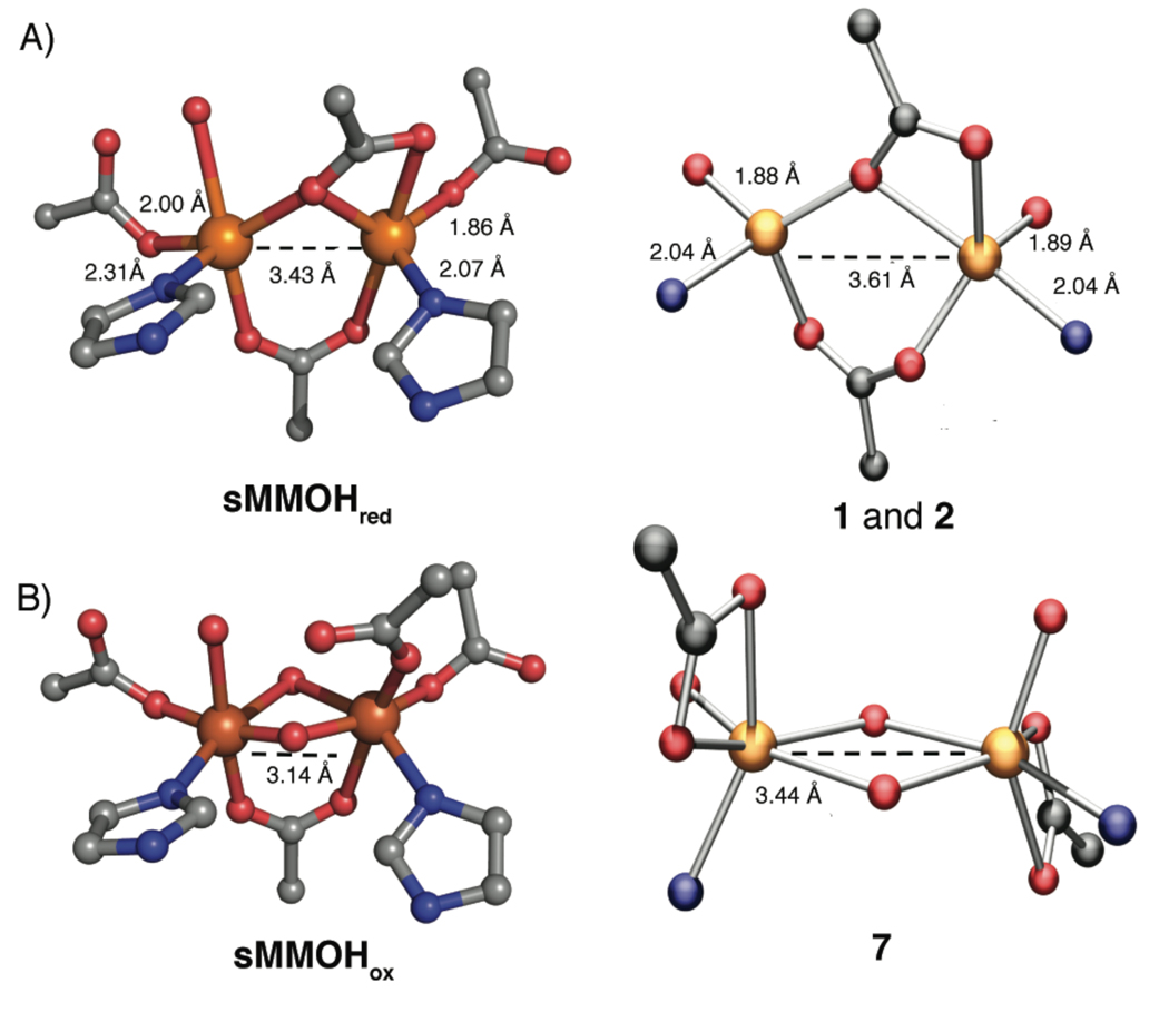
Figure 3.
Depiction of the X-ray crystal structures of the diiron sites of sMMOHred (top, left) and sMMOHox (bottom, left). For structural comparison, synthetic complexes that mimic each protein state are shown on its right. Some relevant bond lengths are provided; the distances shown for 1 and 2 are averaged over the two complexes. Color scheme: iron, orange; nitrogen, blue; oxygen, red; carbon, gray.

