Figure 4.

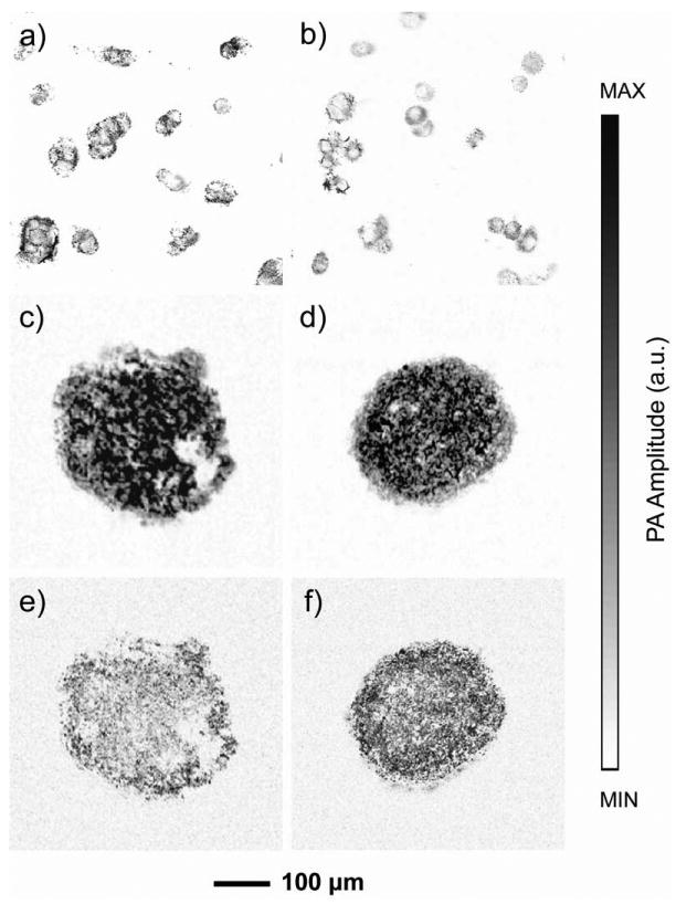
PAM images of a) RW4 mouse embryonic stem cells and b) SK-BR-3 breast cancer cells incubated with MTT for 3 h. In a parallel experiment, the cells were washed with PBS to remove MTT and fresh medium was added after the same MTT staining procedure to validate the non-invasive nature of the staining method. The cells were further cultured for 36 h to ensure complete exocytosis of the MTT formazan crystals. The cells were then induced to form characteristic cell aggregates, i.e., embryoid bodies in the case of RW4 mESCs or multicellular tumor spheroids in the case of SK-BR-3 breast cancer cells, and then stained with MTT again for PAM. c, d) PAM images of an embryoid body and a multicellular tumor spheroid after MTT staining. e, f) MAP images taken from the middle planes (60 μm in thickness) of the spheroids shown in (c) and (d), respectively.
