Figure 6.
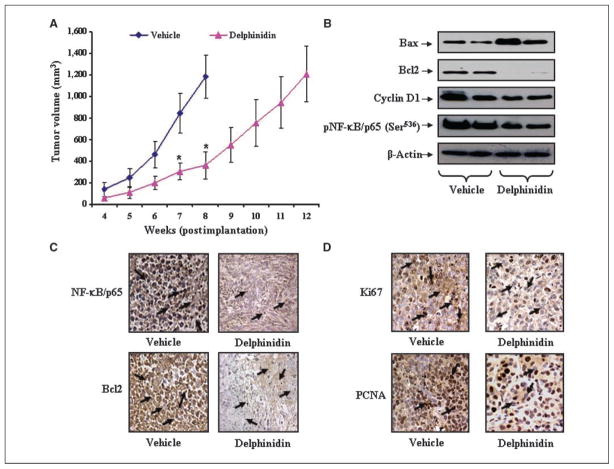
Effect of delphinidin administration on tumorigenecity of PC3 cells and the expression levels of Bax, Bcl2, NF-κB/p65, and known proliferation markers, Ki67 and PCNA, under in vivo conditions. A, average tumor volume of vehicle- or delphinidin-treated animals was plotted over weeks as detailed in Materials and Methods. Points, mean of 12 tumors; bars, SD. P < 0.01, versus vehicle-treated animals. B, protein levels of Bax, Bcl2, cyclin D1, and phospho-NF-κB/p659(Ser536) as determined by immunoblot analysis in pooled tumors excised from mice treated with vehicle or delphinidin. Equal loading of protein was confirmed by stripping and reprobing the blots with β-actin antibody. C and D, representative photomicrographs (magnification, ×200) showing immunohistochemical staining for NF-κB/p65, Bcl2, Ki67, and PCNA in tumor sections of vehicle- or delphinidin-treated mice harvested at 8 wk. Arrows, regions exhibiting immunoreactivity.

