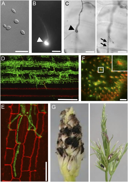
Figure 1.
Morphological stages of S. reilianum outside, on, and in maize cv Gaspe Flint. A, Axenically grown haploid sporidia of S. reilianum. Bar = 15 μm. B, On the plant surface, dikaryotic hyphae of S. reilianum form appressoria for plant penetration at 1 d after infection. An appressorium (arrowhead) is visualized here by fluorescence microscopy after Calcofluor staining. Bar = 15 μm. C, Appressoria of S. reilianum penetrate the leaf surface. From the appressorium (arrowhead; left panel), a hypha penetrates the underlying epidermal tissue (arrows; right panel). Images in the left and right panels are of different focal planes visualized by bright-field microscopy after staining with Chlorazole Black E. Bars = 10 μm. D, In the plant leaf, fungal hyphae proliferate along leaf vascular bundles. The image shows a Z-stack of WGA-Alexa Fluor-stained (green; fungal hyphae) and propidium iodide-stained (red; plant cells) samples visualized by confocal microscopy. Bar = 300 μm. E, Closeup of fungal hyphae colonizing bundle sheath cells. Bar = 50 μm. F, In the nodes of the plant, fungal hyphae (green) surround the plant vascular bundles (red). A cross-section stained with WGA-Alexa Fluor and propidium iodide was visualized by confocal microscopy. Bar = 500 μm. G, Spores of S. reilianum forming on maize ear (left) and tassel (right).

