Figure 9.
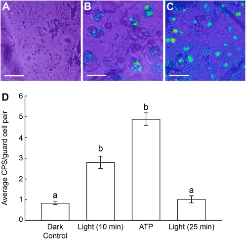
Light treatment of dark-adapted leaves induces the release of ATP in guard cells, as assayed by ecto-luciferase luminescence. A, Background levels of ecto-luciferase luminescence are observed in an epidermal peel from an untreated x-luc9 leaf (dark control). B, An epidermal peel from an x-luc9 leaf treated with 10 min of light shows ecto-luciferase luminescence in guard cells. C, An epidermal peel from an x-luc9 leaf treated with 1 mm ATP in the dark shows ecto-luciferase luminescence in guard cells. Bars = 50 μm for A and B and 100 μm for C. Luminescence levels are represented in pseudocolor (blue, green, yellow, orange, and red, where red represents the highest and blue represents the lowest level of relative intensity). D, Quantification of luciferase activity from a representative data set of an opening experiment. Treatment with 1 mm ATP was done in the dark. Luminescence returned to untreated control levels 25 min after treatment with light. Different letters above the bars indicate mean values that are significantly different from one another as determined by Student’s t test (P < 0.05; n ≥ 15 guard cell pairs). These data are representative of three biological repeats. Error bars represent se.

