Figure 2.
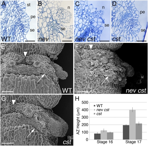
nev cst AZs are disorganized and enlarged. A to D, Longitudinal sections of flowers (stage 17) stained with toluidine blue. The remaining AZ cells of wild-type (WT; A) and cst-2 (D) flowers show coordinated cell expansion, while the floral organs remain attached in nev flowers (B). Although organ abscission is rescued in nev cst-2 flowers (C), the AZ cells have a disordered appearance. The petal (pe), sepal (se), and stamen (st) AZs and nectaries (n) are indicated. Bars = 50 μm. E to G, Scanning electron micrographs of flowers after organ separation (stage 17). Distinct AZs are apparent in wild-type (E) and cst (G) flowers, whereas the AZ regions of nev cst flowers have formed an enlarged, disorganized band of cells at the fruit base (F). In nev cst flowers, the junction between the medial stamen AZs is no longer visible (arrowheads), and the border between the sepal AZ and the floral stem is not clearly defined (arrows). Bars = 500 μm. H, Quantification of AZ size in wild-type and mutant flowers. The distance between the lower border of the sepal AZ and the upper border of the stamen AZ was measured in stage 16 and the first stage 17 flowers (n ≥ 4 per genotype). nev cst flowers contain significantly enlarged AZs after organ shedding compared with wild-type and cst flowers. [See online article for color version of this figure.]

