Figure 2.
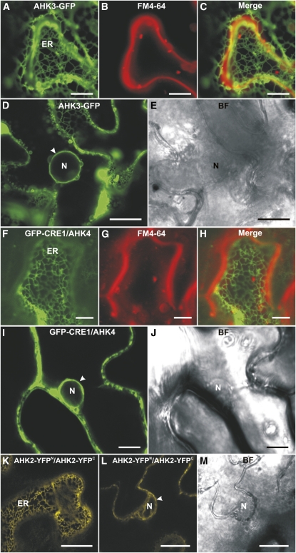
Localization of fluorescent cytokinin receptor fusion proteins in epidermal cells of tobacco leaves. A, ER labeled with AHK3-GFP. B, Staining of the PM with FM4-64 (50 μm). C, Merged image of A and B. D, Localization of AHK3-GFP in the perinuclear space (arrowhead). E, Bright-field (BF) image of D. F, ER labeled with GFP-CRE1/AHK4. G, Staining of the PM with FM4-64 (50 μm). H, Merged image of F and G. I, Localization of GFP-CRE1/AHK4 in the perinuclear space (arrowhead). J, Bright-field image of I. K, BiFC analysis reveals the formation of AHK2-YFPN/AHK2-YFPC homodimers in the ER. L, Homodimerization of AHK2-YFPN and AHK2-YFPC in the perinuclear space (arrowhead). M, Bright-field image of L. N, Nucleus. Bars = 7.5 μm in F to H; 10 μm in A to C, I, and J; and 25 μm in D, E, L, and M.

