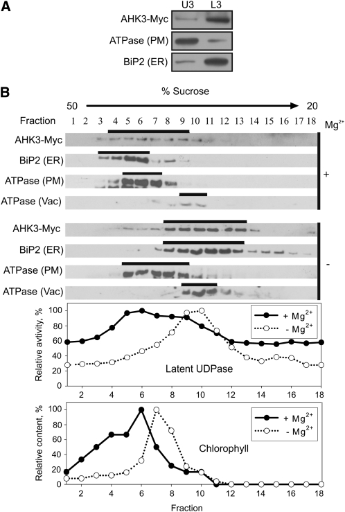
Figure 3.
Localization of AHK3-Myc in fractionated membranes from 6-d-old Arabidopsis seedlings expressing PAHK3:AHK3-Myc. A, Aqueous two-phase partitioning of microsomes. Equal amounts of protein from upper (U3) and lower (L3) phases were separated by SDS-PAGE and subjected to immunoblot analysis with antibodies specific for Myc, H+-ATPase (PM), and BiP2 (ER). B, Microsomal membranes were fractionated on linear 20% to 50% (w/w) Suc gradients in the presence of Mg2+ (+) to stabilize ribosomes at the ER or in the absence of Mg2+ (−) to dissociate ribosomes from the ER. Samples (50–100 μL) of each fraction were analyzed by immunoblot using antibodies specific for Myc, H+-ATPase, BiP2, and V-ATPase (Vac, vacuole membrane marker). Latent UDPase activity was used as an enzymatic marker for the Golgi apparatus, and chlorophyll was used to localize thylakoid membranes. In both cases, the highest measured enzyme activity (A650) and chlorophyll content (A652) were set at 100%.

