Figure 6.
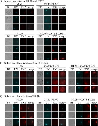
In situ association between HL2b and CAT3 in Arabidopsis cells. Protoplasts were transfected with HL2b, CAT3-FLAG, or HL2b + CAT3-FLAG. BF indicates images observed with bright-field light microscopy. Protoplast nuclei were visualized using Hoechst 33342 staining and UV light (UV). For detection of in situ molecular interaction, the Duolink in situ PLA kit was used. A, Interaction between HL2b and CAT3. Primary antibodies against FLAG and CMV 2b were used to detect CAT3 and 2b, respectively. With fluorescence microscopy, PLA red signals from TX Red (TX2) will theoretically be detected only when there is an in vivo interaction between 2b and CAT3. The images of UV and TX2 were merged (merge column) to identify the exact localization of the signals. B, Subcellular localization of CAT3-FLAG in protoplasts. CAT3 was detected using anti-FLAG antibodies as red signals of TX2. Note that CAT3 was detected in nuclei when HL2b was expressed with the CAT3 gene, while the protein was dispersed in cells without HL2b. C, Subcellular localization of HL2b in protoplasts. HL2b was detected using anti-2b antibodies, which yielded PLA red signals from TX2. As expected, HL2b was localized in nuclei.

