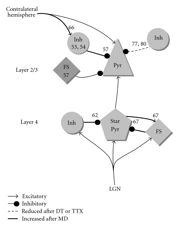
Figure 2.

Documented changes in inhibition in V1 after monocular deprivation (MD), dark treatment (DT) or intraocular TTX injection during the critical period. Numbers correspond to the references that measured the change in the responses or synaptic strength of interneurons. Pyr is pyramidal cell, Star pyr is star pyramid neuron, Inh is interneuron, FS is fast-spiking interneuron. Light gray means shifted towards the open eye after MD. Dark gray is shifted towards the deprived-eye. All studies were done in binocular visual cortex, except for references [67, 77] which were done in monocular cortex and possibly reference [80], which left the exact location within visual cortex unspecified.
