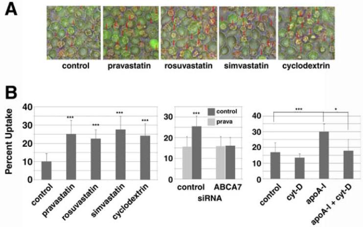
Figure 5. Statins enhance phagocytosis of apoptosis cells by ABCA7.
A: J774 cells were subcultured as 5 × 10⁵ cells/well for one day. After overnight incubation with 50 μM pravastatin, 5 μM rosuvastatin, 10 μM simvastatin or 5 mM cyclodextrin, a quantitative phagocytosis assay for apoptotic Jurkat cells was performed, being shown as representative photos with phagocytosis indicated by red arrows. B: Left, the results of quantitative analysis for the experiments of Figure 5A. Center, knock-down of ABCA7 and phagocytosis of apoptotic cells. ABCA7-specific siRNA was transfected to J774 cells at a density of 8 × 10⁶ cells/cuvette. The cells were subcultured in a 24-well tray as 1 × 10⁶ cells/well. After overnight incubation with and without 50 μM pravastatin, quantitative phagocytosis assay for apoptotic cells was performed. Right, enhancement of phagocytosis of apoptosis cells by apoAI and its inhibition by cytochalasin D. J774 cells were subcultured as 5 × 10⁵ cells/well for one day. After overnight incubation with 10 μg/ml apoAI, 10 μM cytochalasin D, and both, quantitative phagocytosis assay for apototic cells was performed. The data represent the mean ± SD for six samples. Statistical significance is indicated as * for P < 0.05 and *** for P < 0.001.

