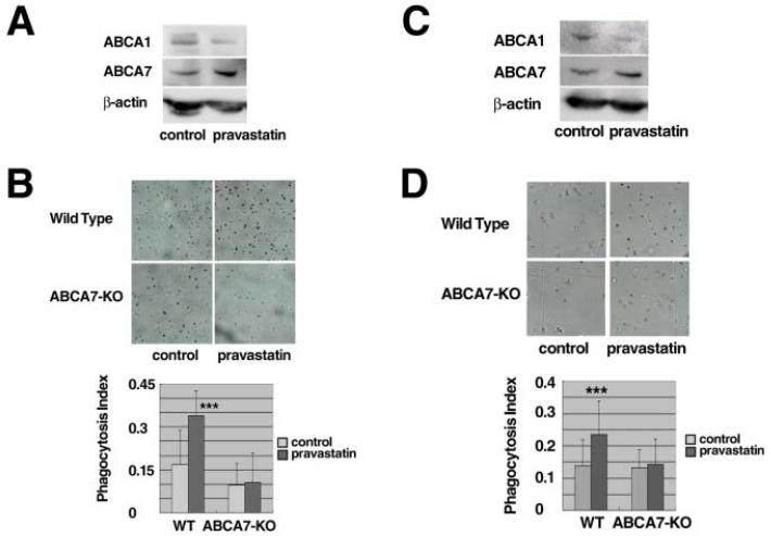
Figure 6. Pravastatin enhances phagocytosis of peritoneal cells in vivo.
A, C: Wild type mice were treated with and without pravastatin by peritoneal (A) and subcutaneous (C) injection as described in the text. Peritoneal cells were collected from the mice, two mice for one sample, and analyzed for proteins by Western blotting immediately after collection. B, D: Phagocytic activity was measured directly in the peritoneal cavity of the mice in vivo. Diluted carbon ink with or without pravastatin was given intraperitoneally (B) or subcutaneously (D). After overnight starvation, peritoneal macrophages were recovered and over 100 cells were counted for calculation of the phagocytosis index as the relative number of cells that engulfed carbon ink particles. Data represents mean ± SD of n = 4 for wild type and ABCA7-knockout mice. Statistical significance is indicated as *** for P < 0.001 against each control.

