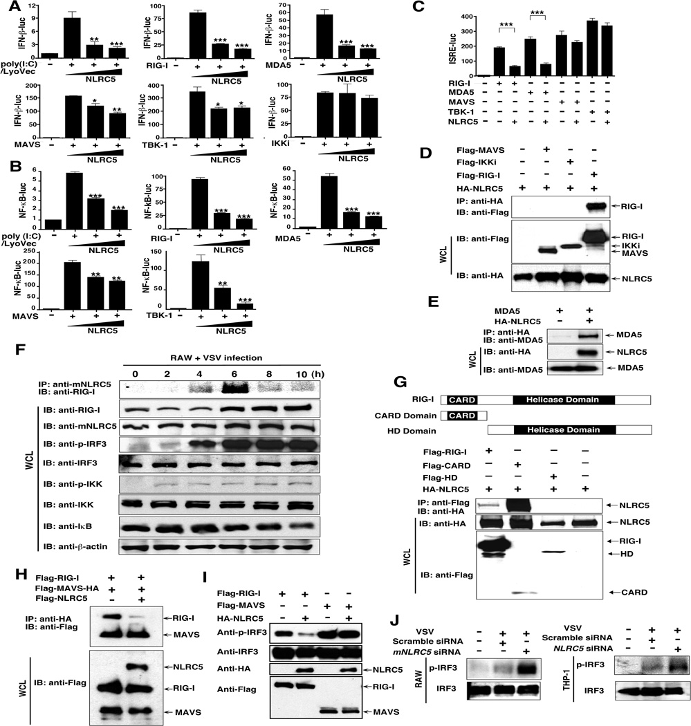
Figure 6. NLRC5 negatively regulates IFN-β activation by inhibiting RIG-I and MDA5 function.
(A–C): 293T cells were transfected with NF-κB-luc, INF-β-luc or ISRE-luc, NLRC5 plus poly(I:C)/Lyovec, RIG-I, MDA5, MAVS, TBK1 or IKKi plasmids and analyzed for INF-β or ISRE luciferase activity. Values are means ± SEM of three independent experiments.
(D) 293T cells were transfected with HA-NLRC5 plus RIG-I, MAVS or IKKi. After immunoprecipitation with anti-HA beads, specific proteins were analyzed by Western blot with anti-Flag.
(E) 293T cells were transfected with MDA5 with or without HA-NLRC5. After immunoprecipitation with anti-HA beads, specific proteins were analyzed by Western blot with anti-MDA5.
(F) RAW264.7 cells were infected with VSV-eGFP, and cell extracts were harvested at different time points, immunoprecipitated with anti-mNLRC5 antibody and analyzed by Western blot with anti-RIG-I.
(G) NLRC5 binds to the CARD domain of RIG-I. 293T cells were transfected with HA-NLRC5 plus Flag-RIG-I, Flag-RIG-I CARD domain and Helicase domain (HD). After immunoprecipitation with anti-Flag beads, specific proteins were analyzed by Western blot with anti-HA.
(H) NLRC5 competitively binds to RIG-I with MAVS. HEK293T cells were transfected with Flag-MAVS-HA plus Flag-RIG-1, or Flag-NLRC5. After immunoprecipitation with anti-HA beads, specific proteins were analyzed by Western blot with anti-Flag.
(I) 293T cells were transfected with Flag-RIG-I and Flag-MAVS, with or without HA-NLRC5, and used for Western blot analysis with anti-phospho-IRF3 and IRF3 antibodies.
(J) RAW264.7 and THP-1 cells were transfected with NLRC5/mNLRC5-specific siRNA or scrambled siRNA, respectively, and then infected with VSV-eGFP. The cell extracts were harvested for Western blot with anti-phospho-IRF3 and IRF3 antibodies.
See also Fig. S6.

