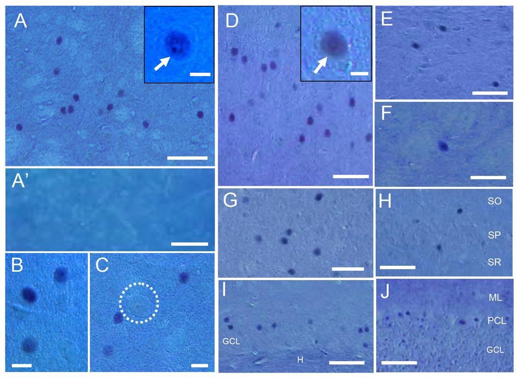
Fig. 2. Immunohistochemical staining of N-TAF1 under Nomarski optics in multiple brain regions.
(A–C) Striatal section stained for N-TAF1 shows that N-TAF1+ nuclei are in medium-sized cells. It is also shown that large (giant)-sized cells (C, dashed open circle) are devoid of N-TAF1 labeling. A high-power image of the neuron possessing strong N-TAF1 labeling in its nucleus and nucleoli (arrow) is shown in the inset in A. Note that no N-TAF1+ nuclei are found in a striatal section incubated with the anti-N-TAF1 antibody (mAb-3A11F) preabsorbed with excess of KLH-conjugated N-TAF1-peptide (A’).
(D–J) Distribution of N-TAF1+ nuclei in non-striatal brain regions that include the cerebral cortex (D), substantia nigra (E), globus pallidus (F), thalamus (G), hippocampus proper (H) and dentate gyrus (I), and cerebellum (J). A high-power image of cortical neuron possessing strong N-TAF1 labeling in its nucleus is shown in the inset in D.
Scale bars: A, A’ and D–J, 100 µm; B, C, 20 µm; insets in A and D, 10 µm.
SO, stratum oriens; SP, stratum pyramidale; SR, stratum radiatum, GCL, granule cell layer; H, hilus dentata; ML, molecular layer; PCL, Purkinje cell layer.

