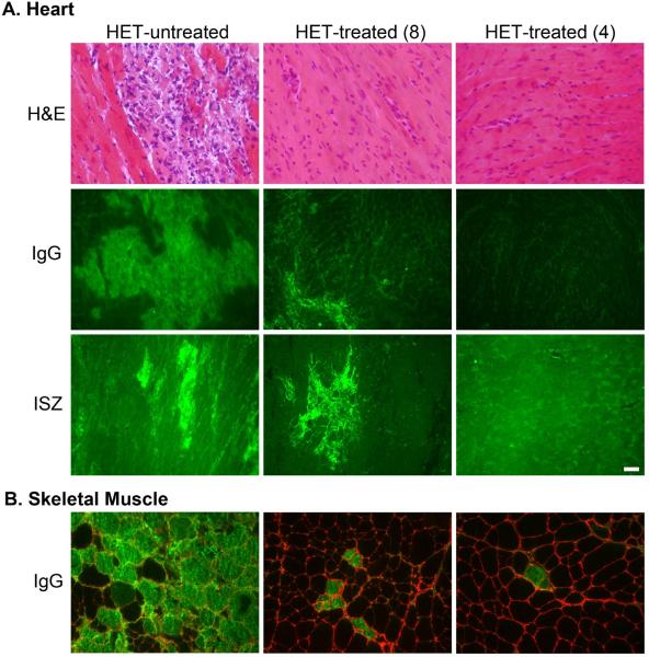
Figure 3.
Drug treatment improves histological parameters of heart and skeletal muscles. A) Hematoxylin and Eosin (H&E)-stained left ventricular sections show the cardiac damage prevalent throughout het-untreated hearts that is almost completely prevented in both treatment groups. Intracellular localization of mouse IgG (green) indicates damaged myocardium that is significantly attenuated in het-treated-8 and even further improved in het-treated-4 hearts. Gelatinase in situ zymography (ISZ) shows the combined activity of matrix metalloproteinases 2 and 9 (bright green), indicative of ventricular remodeling, that is also attenuated in the het-treated-8 hearts, and almost entirely prevented in the het-treated-4 hearts. B) IgG localization (green) in quadriceps skeletal muscle sections indicates a profound and significant reduction of ongoing myofiber damage in the het-treated-4 group, with intermediate effects in the het-treated-8 group compared to untreated hets. Localization of Collagen I (red) in the matrix surrounding individual muscle fibers is shown to demonstrate the intracellular localization of the IgG staining. Bar = 50 μm.

