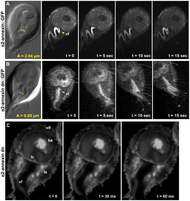
Figure 3. Overexpression of a dominant negative alpha2-annexin::GFP (D122A, D275A) decreases ventral flagellar waveform amplitude with only slight decreases in attachment.
The role of the ventral flagella in attachment was determined using overexpression of a dominant negative form of the ventral flagella-associated alpha2-annexin (see Methods). DIC microscopy was used to measure the amplitude (A) of the ventral flagellar waveform (vf). Fluorescence microscopy was used to determine the localization of the GFP-tagged alpha2-annexin. Panel (A) shows that alpha2-annexin localizes primarily to the ventral flagella and diffusely to the plasma membrane of the ventral disc, yet GFP-tagging does not affect flagellar beat frequency or amplitude. (B) Overexpression of the dominant negative alpha2-annexin (D122A, D275A), or alpha2-annexin dn, results in a ventral flagellar (vf) beat with significantly decreased amplitude (n = 25; see also Video S3). Panel (C) shows trophozoite surface contacts using cell membrane stained trophozoites using TIRFM. As compared to the standard in Figure 1, proper substrate surface contacts are made, including a continuous disc seal (lc), lateral shield (ls) and bare area (ba). The same trophozoite surface contacts and disc perimeter seal are visible despite a diminished flagellar waveform amplitude and asynchrony between the flagella (see also Video S4).

