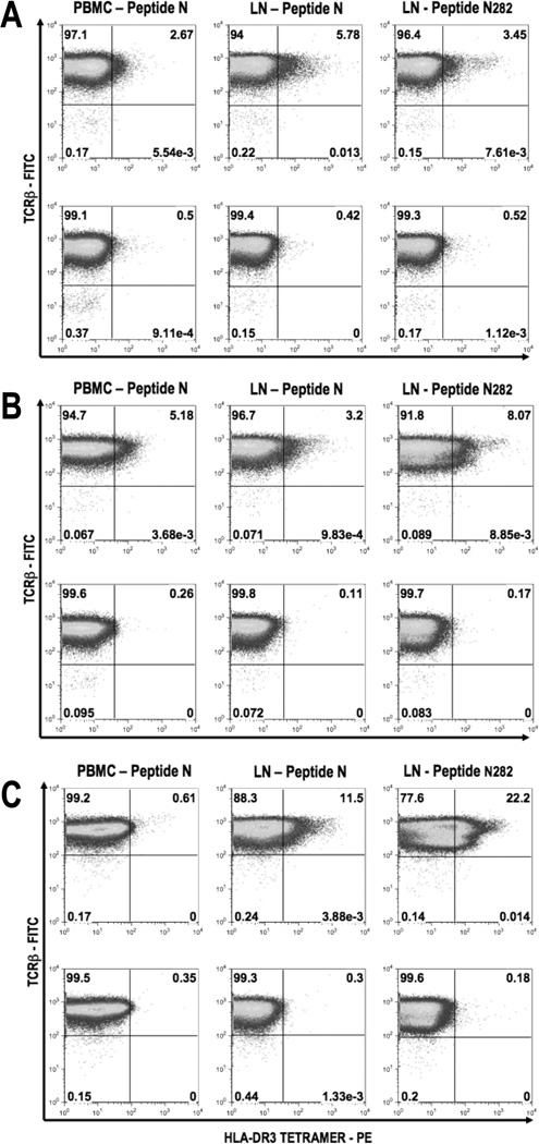
Figure 5. Peptide loaded HLA-DR3 tetramer detect epitope-specific CD4+ T cells from peripheral blood and draining lymph nodes of immunized HLA-DR3(0301) Tg mice.
HLA-DR3 Tg mice were immunized with S-Ag twice as described in Methods. Cells were isolated from either peripheral blood or draining lymph nodes 12 days after the second immunization. PBMC were in-vitro stimulated with peptide N and draining LN cells were stimulated with peptide N or peptide N282 for 48 hours. Cells were expanded in IL-2 containing medium for 7 days and stained using HLA-DR3 tetramer (PE) loaded with peptide N282 as shown in the upper half, or with unloaded HLA-DR3 tetramer (PE) as shown in the lower half of each panel, at 37°C for one hour followed by anti-CD4 (APC) and anti-TCRβ (FITC). Cells were also stained with 7AAD to exclude the dead cells from analysis. Percentage of tetramer+ TCRβ+ cells are shown after gating CD4+ 7-AAD− cells after the first round (A), second round (B), and third round (C) of stimulation and expansion. Data shown is from one representative experiment of two.

