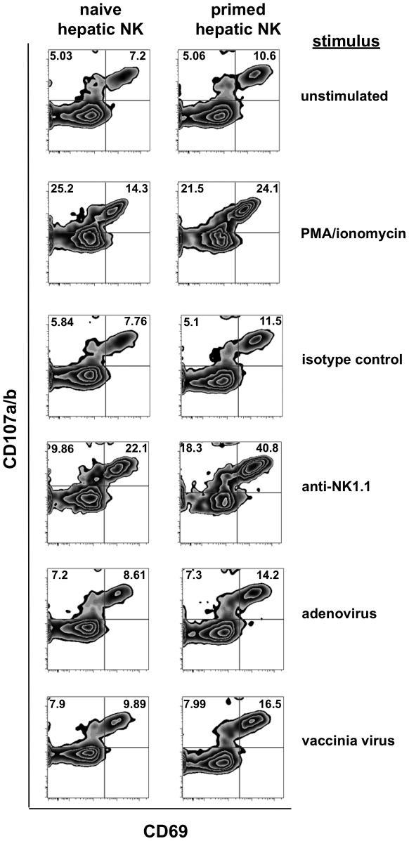
Figure 3. Intracellular cytokine staining of previously primed NK cells demonstrates enhanced activation following in vitro stimulation.
Lymphocytes and NK cells isolated from collagenase-dissociated liver preparations from naïve and vaccinia virus-infected mice were cultured in vitro with the indicated stimuli for 6 hours prior to evaluation for NK cell activation (expression of CD69) and degranulation (expression of CD107) in response to the stimulation. Stimuli include plate-bound isotype control Ig, the anti-NK1.1 monoclonal antibody PK-136, formaldehyde-fixed adenovirus (2×10⁶ vp), and formaldehyde-fixed vaccinia virus (2×10⁶ pfu). Plots shown are gated on NK cells (NK1.1+CD3− or DX5+CD3−).

