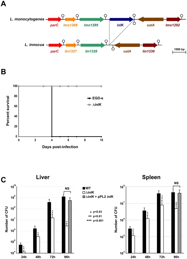
Figure 1. InlK is a virulence factor of L. monocytogenes.
A. The inlK gene locus in L. monocytogenes compared with the same genomic region in the related non-pathogenic species L. innocua. The stem and circle represent transcription terminators. B. Kaplan-Meier curve represents the survival of BALB/c mice over time. Four BALB/c mice in each experimental group were infected i.v with 104 L. monocytogenes wild-type (EGD-e) or ΔinlK mutant. C. The L. monocytogenes EGD-e wild-type strain (WT), the ΔinlK mutant (ΔinlK) and the complemented strain (ΔinlK+pPL2 inlK) 104 CFU were inoculated i.v into BALB/c mice. Animals were euthanized 24 h, 48 h, 72 h or 96 h after infection and organs were recovered, homogenized, and homogenates serially plated on BHI. The number of bacteria able to colonize liver (left panel) and spleen (right panel) is expressed as log10 CFU. Four animals per bacterial strain, per time points and per experiment were used. Statistical analyses were performed on the results of 3 independent experiments using the Student t test. P values of <0.05 were considered statistically different and are labeled here as *.

