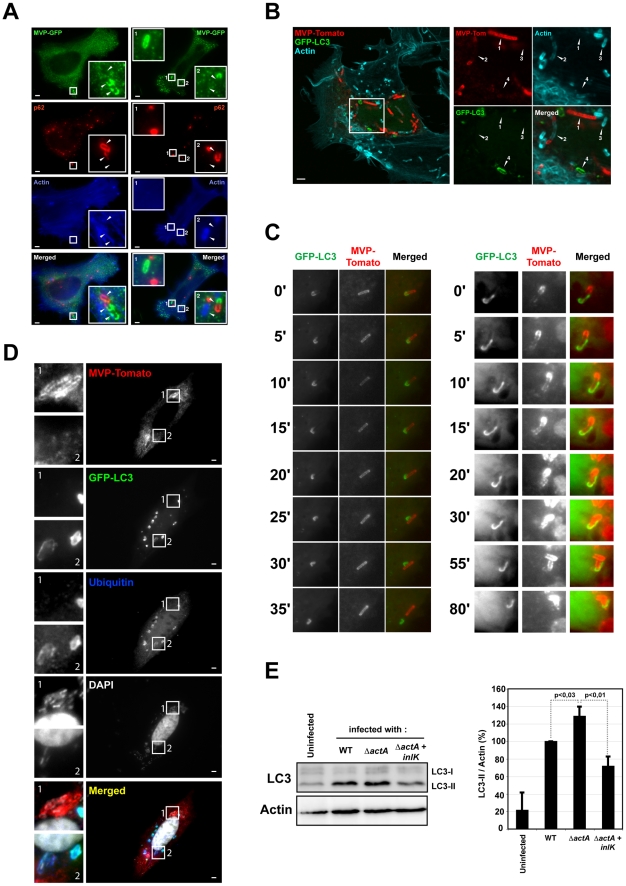
Figure 5. MVP impairs the recruitment of autophagy markers.
A. Impaired recruitment of p62 to MVP positive Listeria. HeLa cells were transfected with MVP-GFP (green), infected with InlK over-expressing Listeria (ΔinlK+pPRT-inlK) for 4 h, fixed for fluorescence light microscopy, and stained with phalloidin (blue) and anti-p62 antibody (red). Inset regions are magnified. Arrows indicate independent bacteria The scale bar represents 1 µm. The vast majority of MVP-positive bacteria were completely devoid of anti-p62 labeling (95.1±2.0%; mean ± SEM from n = 3 experiments) but 4.9±2.0% (mean ± SEM from n = 3 experiments) were stained at one pole with MVP and at the other pole with p62. B. Impaired recruitment of GFP-LC3 on MVP positive Listeria. HeLa cells were transfected with MVP-tomato (red) and GFP-LC3 (green), infected with InlK over-expressing Listeria (ΔinlK+pPRT-inlK) for 4 h, fixed for fluorescence light microscopy, and stained with phalloidin (blue). Inset regions are magnified. The scale bar represents 1 µm. MVP and/or actin positive bacteria were never recognized by GFP-LC3. Arrows point to bacteria at different steps of the infection process: 1) InlK over-expressing bacterium is totally covered by MVP; 2) bacterium is partially labeled with MVP (at the poles) and actin (at the center); 3) bacterium is completely covered by actin; 4) bacterium is enclosed in an GFP-LC3 positive autophagosome. C. Kinetics of autophagy escape for MVP positive Listeria. Jeg3 cells were transfected with MVP-tomato (red) and GFP-LC3 (green), infected with InlK over-expressing Listeria (ΔinlK+pPRT-inlK) for 4 h, and prepared for real-time video microscopy. Image series were collected every 5 min for 2 h. Time is indicated along the Y axis. The left panel shows that the GFP-LC3 membranous aggregate detaches from the MVP positive Listeria. The entire image sequence can be viewed as Video S2. The right panel shows that the GFP-LC3 membranous aggregate on MVP positive bacteria does not lead to an autophagosome formation, whereas those bacteria efficiently divided. The entire image sequence can be viewed as Video S3. D. Impaired recruitment of GFP-LC3 and ubiquitin to MVP positive ΔactA Listeria. HeLa cells were transfected with MVP-tomato (red) and GFP-LC3 (green), infected with InlK over-expressing ΔactA (ΔactA+pADc-inlK) for 4 h, fixed for fluorescence light microscopy, and stained with anti-ubiquitin antibody (blue) and DAPI (white). Inset regions are magnified. The scale bar represents 1 µm. E. LC3 levels in infected RAW 267.4 macrophages. Left panel: RAW 267.4 macrophages were infected with L. monocytogenes EGD (WT), ΔactA or ΔactA+InlK for 6 h. Cell total lysates were immunobloted for LC3 and actin. Western blot is representative from 3 independent experiments. Right panel: Quantification of the relative LC3-II level (mean ± SEM) shown in the left panel. Statistical analyses were performed on the results of 3 independent experiments using the Student's t test. P values of <0.05 were considered statistically different.

