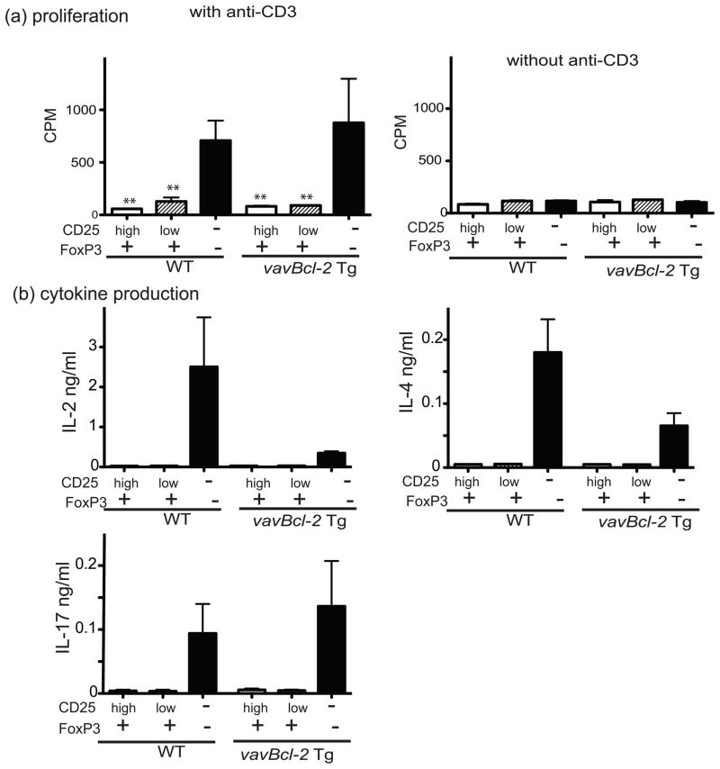
Fig. 5. CD25lowFoxP3+ cells are hypo-responsive to TCR stimulation.
(a) T cell proliferation: Distinct CD4+ T cell subpopulations from lymph nodes of FoxP3GFPKI or FoxP3GFPKI/vavBcl-2 Tg mice were sorted according to the levels of FoxP3-GFP and CD25 expression. These T cell subsets (2x10⁴ cells per well) were cultured in 96-well round-bottomed plates with 5 μg/mL anti-CD3 plus 2 μg/mL anti-CD28 Abs in the presence of 8x10⁴γ-irradiated and T cell depleted spleen cells (used as antigen presenting cells). The mean cpm +/− SD from 3–5 replicates per culture condition is shown. **P<0.01 compared to GFP-CD25−CD4+ lymph node cells. Three experiments were performed with similar results. (b) Cytokine production: supernatants from the above cultures harvested after 3 days of incubation were examined for cytokine content. Data represent the mean +/− SEM from three replicate cultures per cell type and genotype. Three independent experiments were performed and produced similar results.

