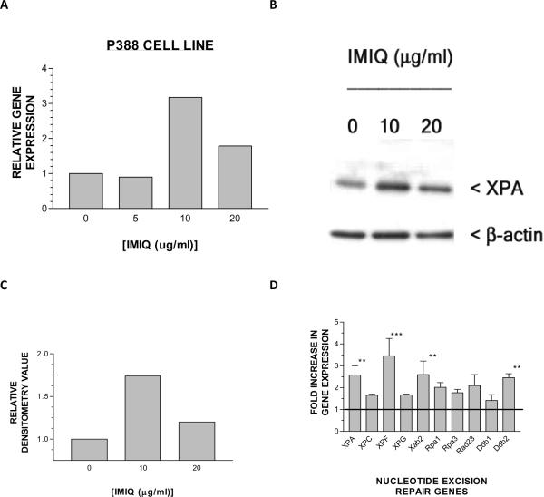
Figure 1. Imiquimod enhances gene expression and nuclear localization of the DNA repair enzyme, XPA in bone marrow derived cells.
(A)The P388 cell line was incubated with medium alone, or medium containing the indicated doses of imiquimod, for 6 hours, and then gene expression was assayed using qPCR analysis (single representative experiment, see methods). (B) Western blotting of nuclear XPA (approximately 38kD) 24 hours after P388 was incubated with medium alone, or medium containing the indicated doses of imiquimod. (C) Densitometry scanning of normalized XPA/β-actin indicated that there was close to a two-fold increase in nuclear XPA protein in imiquimod treated cells. (D) Fold change expression of multiple DNA repair genes in P388 cells cultured with 10 μg/ml of imiquimod relative to cells cultured in medium alone (not shown, but depicted as horizontal line) (The data in this experiment represent the mean and standard deviation of three different gene expression experiments; **p < 0.01; ***p < 0.001, ANOVA).

