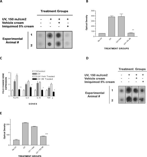
Figure 7. Topical applications of imiquimod cream decrease the amount of DNA damage found in local lymph nodes and immune suppressive cytokines after a single cutaneous UVL exposure.
Experimental animals were un-irradiated, or irradiated with a single dose of 150mJ/cm2 of UVB on the dorsal surface of the pinna of the ear. The UVL irradiated mice were either untreated, treated with topical applications of vehicle (placebo) cream or 5% imiquimod cream for two consecutive days prior to the UVL exposure. Twenty four hours after the single UVL exposure, the cervical lymph nodes were dissected from all four groups of animals and DNA was extracted as described in the methods. (A) DNA dot blot, showing decreased CBPD only in imiquimod treated mice compared to the UV irradiated, untreated or the vehicle treated groups of mice, but similar to the mice not exposed to UV. (B) Summary of densitometry scanning (data represent mean +/− SD of the two depicted animals; **p < 0.01, ANOVA). (C) qPCR analysis of immune suppressive gene expression (IL-4, IL-10, TGF-β and FoxP3) in local lymph nodes, using experimental protocol as described above. These data represent data from two animals from each experimental group (data depicted represent mean +/− SD; ** p<0.01, highly significant; * p <0.05 compared to vehicle control, significant, ANOVA); this experiment was repeated three times, and found to be reproducible. (D) DNA dot blot depicting CBPD from lymph nodes derived from IL-12 knockout mice (same protocol used for wild type mice as described above). (E) Summary of densitometry scanning (data represent mean +/− SD of the two depicted animals; ***p < 0.001, ANOVA).

