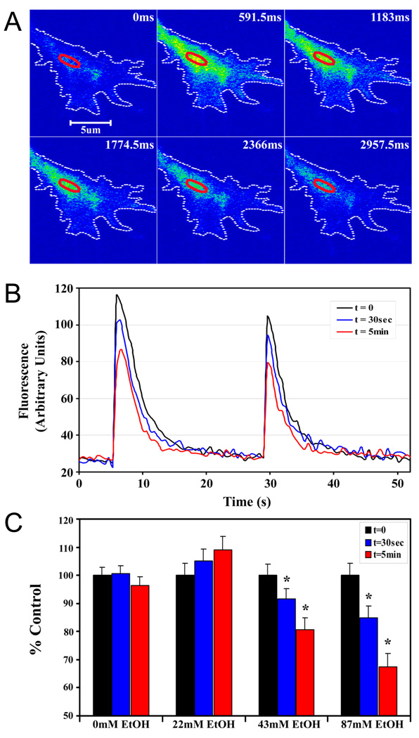
Figure 5. Acute ethanol inhibits depolarization-evoked calcium signals in axonal growth cones.
A. Representative pseudocolor fluorescence images of a KCl-induced Ca2+ in the axonal growth cone of a hippocampal pyramidal neuron 24 h after plating. B. Traces of Ca2+ in a region of interest in the palm of the same growth cone, when evoked by KCl pulses of 500 msec (left) and 250 msec (right), before (t = 0; black line), and at 30 sec (blue line) and 5 min (red line) after bath application of 87 mM ethanol. Ethanol inhibition of the peak fluorescence intensity increases with time. Ethanol does not alter the graded response to pulse duration and Ca2+ returns to baseline between pulses. C. Histogram plot of peak amplitudes of KCl-evoked calcium transients in axonal growth cones before, and at 30 sec and 5 min after the addition of 22, 43 or 87 mM ethanol. Data are the mean ± SEM peak fluorescence expressed as a percent of control transients evoked by the 500 msec pulse. The inhibitory effect of ethanol is dose- and time-dependent. * P < 0.01, ** P < 0.001; repeated measures ANOVA with Newman-Keuls posthoc test.

