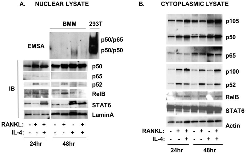
Figure 2. Effect of RANKL and IL-4 on the slow activation of NF-κB.
Primary BMM were cultured in the presence or absence of RANKL (150 ng/ml) or IL-4 (10 ng/ml) for various times as indicated. Nuclear and cytoplasmic lysates were prepared as described in the Methods section. A. Top panel. Nuclear lysates were incubated with the biotin-labeled κB oligo and analyzed by EMSA assay as described in the Methods section. Total lysates of 293T cells transfected with p50 and p65 were run simultaneously as reference. Bottom panel, Nuclear lysates were analyzed by western blotting using anti-p50, anti-p65, anti-p52, anti-RelB, anti-STAT6, or anti-lamin A antibodies. B. Cytoplasmic lysates were analyzed by western blot using anti-p105/p50, anti-p65, anti-p100/p52, anti-RelB, anti-STAT6, or anti-actin antibodies.

