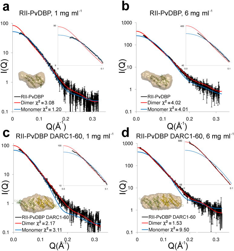
Figure 2.
DARC binding drives dimerization of RII-PvDBP. Experimental (black) and theoretical SAXS plots for the monomer (blue) and dimer (red) at different concentrations. An expanded plot of the low-angle data (0 < Q < 0.1) that clearly delineates oligomeric state is shown in the top right insert. Ab initio reconstructions are overlayed on structures (bottom left insert) with monomers colored in green and yellow and molecular envelopes in sand. (a) RII-PvDBP at 1 mg ml−1. (b) RII-PvDBP at 6 mg ml−1. (c) RII-PvDBP–DARC1–60 at 1 mg ml−1. (d) RII-PvDBP–DARC1–60 at 6 mg ml−1.

