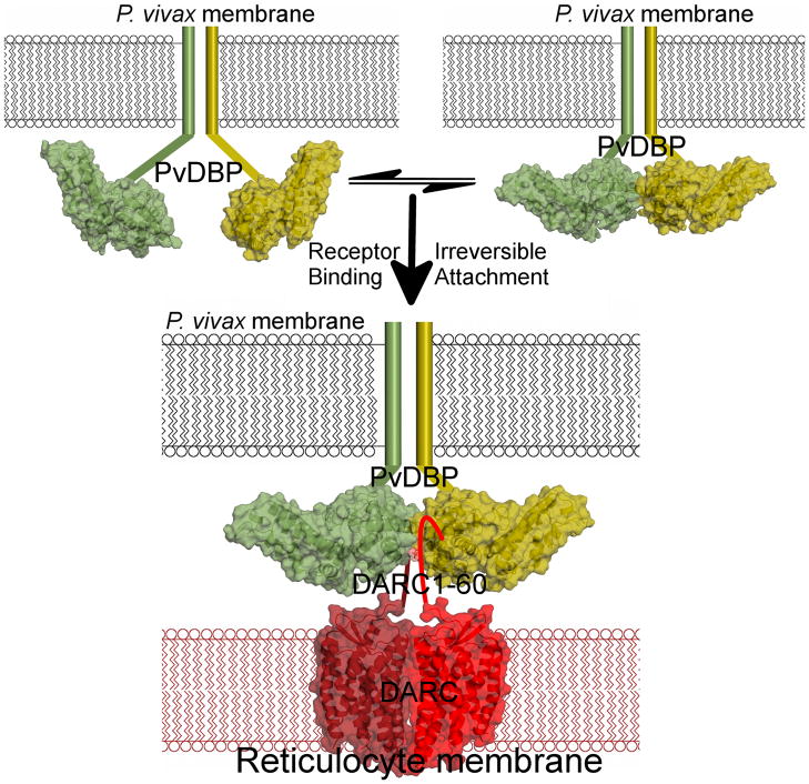
Figure 5.
PvDBP binds DARC via a model of receptor-mediated ligand-dimerization. PvDBP exists as an equilibrium of monomers and dimers that is shifted to dimerization upon receptor-binding. RII-PvDBP monomers are in green/yellow. The P. vivax membrane is in black and the reticulocyte membrane is in red. Flat lines represent portions of PvDBP not in the crystal structure. The DARC homodimer is represented by the crystal structure of a related GPCR, CXCR4’s, homodimeric membrane spanning region40, in dark/light red. DARC1–60 is shown as a flat line. Two PvDBP molecules bind two DARC molecules as indicated by our stoichiometry measurements.

