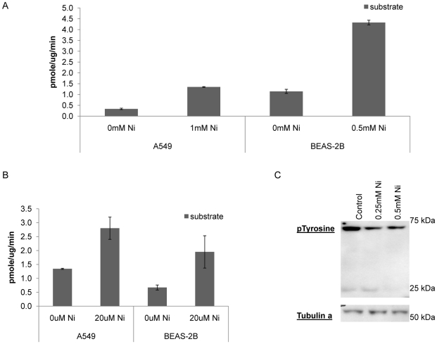
Figure 3. Increases protein tyrosine phosphatase activity following nickel treatment.
Free phosphates which were generated by dephosphorylation reaction of the substrate were measured. (A) PTP activity was up regulated in BEAS-2B cells and A549 cells after 24 h nickel treatment. (B) Un-treated control cells showed increased phosphatase activity when nickel was added to cell extracts (C) By western analysis we demonstrated that the level of phosphorylated tyrosines in proteins was reduced in a concentration dependent manner after nickel treatment. This finding correlates with the finding that the phosphatase activity was increased after nickel exposure.

