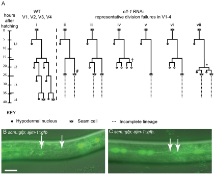
Figure 4. Reduction of elt-1 function causes V lineage division defects.
(A) Lineage traces of WT and elt-1(RNAi) hermaphrodites. In elt-1(RNAi) worms, seam cells often fail to divide but remain intact and retain their stereotypical seam cell ‘eye’ shape. One or more divisions may be missed, for example in lineage trace ii (#). There was no obvious bias for the V cell involved. Alternatively, certain V lineage seam cells sometimes undergo the symmetrical and asymmetrical divisions during L2, but significantly later relative to both other V cells and to the time of moulting. For example, whereas Vn.p cells should divide first symmetrically and then asymmetrically at the start of L2, these divisions have been observed to occur just before the L3 moult in elt-1(RNAi) animals, around 8 hours after they should have occurred (* in lineage trace vii). Similarly, the L2 symmetric and asymmetric divisions in V1-4 should take place slightly ahead of the comparable V6 divisions in WT worms. elt-1 RNAi, however, often delays these divisions such that they occur after V6p has divided symmetrically and then asymmetrically in L2. In addition to these proliferation defects, aberrant fate changes at division were also occasionally observed (approximately 10% of defects observed); instead of the division producing two seam cells or one seam daughter and one hypodermal daughter, both cells produced adopted the hypodermal fate, becoming smaller and rounder than normal seam cells, losing expression of the scm::gfp marker and failing to divide further (e.g. † marked on trace iv). This defect was also observed in rnt-1 and bro-1 animals [10]. Designation of the seam fate was based on cell shape, position and division potential, where possible, not scm::gfp expression. In no worms were L1 division defects observed. (B) and (C) elt-1(RNAi) animals carrying the scm::gfp and ajm-1::gfp transgenes. Seam cells often lose scm::gfp expression (white arrows). 52% of elt-1 (RNAi) worms were found to have one or more cells bounded by AJM-1::GFP but lacking SCM::GFP (n = 61) compared to 5% in control worms, fed on HT115 bacteria containing the empty L4440 RNAi feeding vector (n = 62). As can be seen, these cells retain their AJM-1::GFP boundary and seam morphology and were not observed to fuse with hyp7. The loss of SCM::GFP appears to be independent of the cells' stage in the cell cycle and can happen before (B) or after (C) division. Scale bar, 25 µm.

