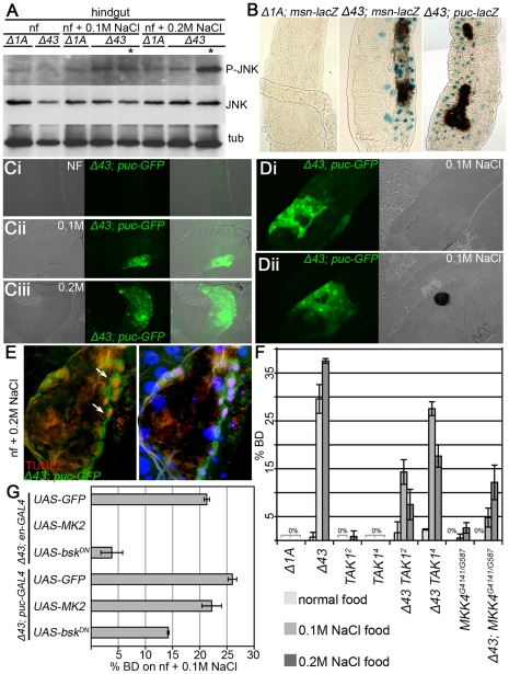
Figure 6. JNK activation leads to EC apoptosis in MK2 deficient hindguts.
(A) JNK phosphorylation is strongly increased in hindguts of MK2 mutant larvae with BDs reared on 0.2 M NaCl food. Lanes containing lysates of larvae with BDs are marked by asterisks. (B) Reporters of JNK signalling activity (msn-lacZ and puc-lacZ) in wild-type (Δ1A) and MK2 mutant (Δ43) larvae reared on 0.2 M NaCl. In MK2 mutants, the JNK activity reporters get induced, especially close to the BDs. (C) Activation of puc-GFP in Δ43 mutants prior to BD appearance. Whereas no activation of puc-GFP occurs on normal food (Ci), fields of increased puc-GFP appear in the hindgut epithelium on 0.1 M NaCl (Cii) and 0.2 M NaCl (Ciii). The area of the patches and the number of affected ECs correlate with the strength of the stress. (D) puc-GFP induction occurs prior to the melanisation process. Hindgut of an MK2 mutant larva at the initiation of a BD (Di) and of an MK2 mutant larva after BD appearance (Dii), both reared on 0.1 M NaCl food. (E) In Δ43 mutants on 0.2 M NaCl, highest puc-GFP (green) and TUNEL staining (red) co-localise in the BD border region (white arrows). (F) Removing two copies of TAK1 or of MKK4 partially suppresses the BD phenotype of MK2 mutants. (G) Overexpression of MK2 and of BskDN in the dorsal hindgut (by means of en-GAL4) rescues the BD phenotype of MK2 mutants on 0.1 M NaCl food. Whereas re-expression of MK2 using puc-GAL4 does not rescue the BD phenotype on 0.1 M NaCl food, downregulation of JNK signalling using UAS-bskDN in combination with the puc-GAL4 driver results in a substantial suppression of the BD phenotype.

