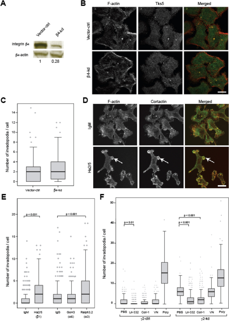
Figure 2. Invadopodia number is increased by blocking integrin β1 but not β4.
(A) Western blotting confirms that β4 was knocked down in ITGB4-kd cells by ~70%. (B) Representative images of vector-ctrl and β4-kd cells cultured on glass for 16 h, fixed, and stained with antibodies against TKS-5 (green) and F-actin (red). Yellow in merged images show colocalization of markers. Scale bar = 10 µm. (C) Box-and-whisker plots show number of invadopodia formed per cell. Knockdown of β4 had little effect on invadopodia formation compared to vector-ctrl cells (N=2; p=0.162). (D) Representative images of γ2-ctrl cells cultured on glass for 3 h, treated with Ha2/5 (integrin β1 blocking antibody; 10 µg/ml) or IgM (10 µg/ml) for 16 h, fixed, and stained with antibodies against cortactin (green) and F-actin (red). Yellow in merged images show colocalization of markers. (E) Blocking integrin β1 by Ha2/5 significantly increased number of invadopodia in γ2-ctrl cells (p<0.001). Scale bar is equal to 10 µm and arrows show podsome-like structure. (F) Plots show number of invadopodia formed per cell type plated on either Ln-332 (1 µg/ml), collagen I (Coll-I; 10 µg/ml), vitronectin (VN; 10 µg/ml), or Poly-D-lysine (Poly; 0.1mg/ml), or PBS. Addition of Ln-332 significantly reduced the formation of invadopodia on both cell types, and Coll-I significantly reduced invadopodia formation of γ2-kd cells (N≥3; P<0.001).

