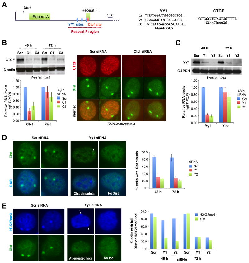
Figure 3. YY1 is required for Xist localization.
A. Map of the proximal 2-kb region of Xist. One CTCF and three putative YY1 binding sites near Repeat F are shown.
B. Western blot, qRT-PCR, and combined Xist RNA FISH/CTCF immunostain 48 hours after Ctcf knockdown using C1 or C3 siRNA. Averages ± SD of three independent experiments shown.
C. YY1 Western blot and Yy1/Xist qRT-PCR after Yy1 knockdown using Y1 or Y2 siRNA. Averages ± SD from 7 independent experiments shown for qRT-PCR. One representative Western blot shown.
D. Xist FISH after Yy1 knockdown. Cells with pinpoint (arrows) or no Xist were scored negative. Averages ± SD from 206–510 nuclei/sample from three independent experiments.
E. H3K27me3 immunostaining (blue) followed by Xist RNA FISH in Yy1-knockdown cells. Two representative patterns shown. Histogram shows counts (n=62–138).

