
Fig. 4.
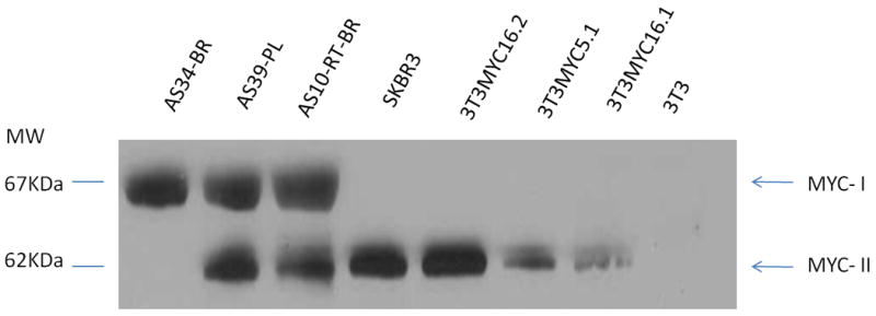
MYC isoforms in AS tumors. Western Blotting with anti-MYC antibody demonstrated that MYC-II (62KDa) expression was abundant in secondary AS tumors (RT-BR, post-radiation breast; PL, post-lymphedema), SKBR3 and MYC-transfected NIH3T3 cells, while not detected in primary AS (BR, primary breast AS). MYC-I (67KDa) was expressed in AS tumors, regardless to RT-exposure, but not in the tested cell lines (SKBR3 and MYC-transfected NIH3T3).