
Figure 4.
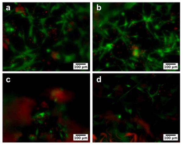
Fluorescence microscopy images showing viability of human mesenchymal stem cells (hMSCs) seeded on membranes formed from 2 wt% HBPA and 1 wt% HA with (a) 0 wt%, (b) 0.1 wt%, (c) 0.25 wt%, and (d) 0.5 wt% heparin at day 5. HMSCs were stained for live (green) and dead (red) cells and appeared to attach and grow on all membranes. The difference in cell density is not a reflection of an effect on proliferation but rather a result of uneven resolution because the membranes are not completely flat and become more wrinkled with increasing heparin concentration. Nonspecific red fluorescence was also seen in membranes containing higher amounts of heparin.