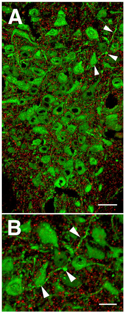
Figure 1. PSD-95 immunolabeling within the mouse DR visualized by array tomography.
A. 3D image of the DR rendered from a stack of 28 ultrathin (70nm) serial sections showing immunolabeling for PSD-95 (red), a marker of excitatory synapses, and tryptophan hydroxylase (TPH) (green), to identify serotonin cells. Tissue sections were immunolabeled with rabbit anti-PSD-95, (1:200, Cell Signaling Technologies) and sheep anti-TPH, (1:200, Millipore). B. Arrowheads in A point to same elements in B at higher magnification, showing the exquisitely discrete labeling of the synaptic marker with total absence of out of focus light achieved in array tomography. Scale bars = 50 um in A, and 20 um in B.

