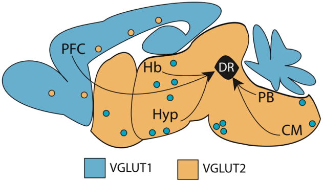
Figure 2. Schematic illustration of major glutamatergic afferents to the DR.

Black arrows indicate brain regions that provide glutamatergic innervation to the DR including the prefrontal cortex (PFC), lateral habenula (Hb) multiple subregions of the hypothalamus (Hyp), the parabrachial nucleus (PB) and areas in the caudal medulla (CM). As a rule, VGLUT1 (light blue) or VGLUT2 (orange) are predominant in cortical and subcortical domains respectively. There are however exceptions to this rule, depicted as polka-dotted colors (Kaneko et al., 2002; Ziegler et al., 2002).
