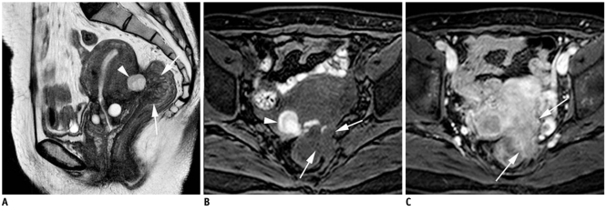
Fig. 10.
Endometriosis that presented as rectal submucosal mass in 46-year-old woman.
Sagittal T2-weighed image (A) shows areas of irregular tissue signal intensity in rectouterine pouch with internal cyst-like structure (arrowhead). Infiltrative rectal wall thickening (arrows) suggests rectal wall involvement. This cyst was identified as hemorrhagic structure by high signal intensity after fat suppression on axial T1-weighted image (arrowhead) (B). Gadolinium enhanced axial T1-weighted image with fat suppression (C) demonstrates heterogeneous enhancing infiltrative mass involving anterior rectal wall and rectouterine pouch (arrows). Lower anterior resection was performed, and endometriosis involvement in submucosa, proper muscle and subserosa of rectum was diagnosed.

