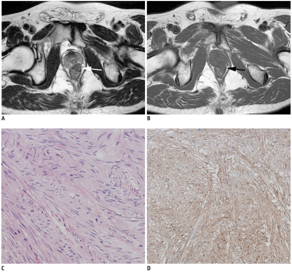
Fig. 5.
Rectal gastrointestinal stromal tumor in 59-year-old woman.
Axial T2-weighted image (A) and T1-weighted image (B) show 1.5-cm submucosal mass (arrows) in left anterior lateral wall of lower rectum. Mass is relatively well-demarcated. After ultralow anterior resection, histopathologic diagnosis was very low-risk gastrointestinal stromal tumor (mitotic rate: 0 per 50 high power fields) based on Hematoxylin & Eosin staining (× 200) (C) and positive c-kit results on immunohistochemistry (D).

