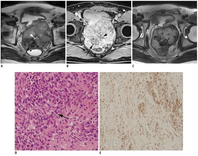
Fig. 6.
Rectal gastrointestinal stromal tumor in 53-year-old man.
Axial T2-weighted image (A) and gadolinium enhanced axial T1-weighted image with fat suppression (B) revealed 9.6-cm multilobulated mass that was possibly arising from anterior wall of rectum. Within mass, dark signal intensities (arrows) due to internal calcification and fluid intensity (arrowheads) due to necrosis are seen. After imatinib treatment, prominent tumor shrinkage is demonstrated on follow-up axial T2-weighted image (C). Mile's operation was performed. Hematoxylin & Eosin staining (× 200) (D) and positive results of c-kit immunohistochemistry (E) revealed diagnosis of high-risk gastrointestinal stromal tumor, in which frequent mitosis (arrow in D; mitotic rate: more than 10 per 50 high power fields) was observed.

