Fig. 1.
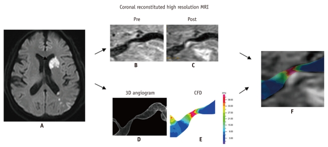
Computational fluid dynamics study in severe M1 stenosis with enhanced plaque seen on high resolution MRI.
Diffusion-weighted image (A) shows perforator and borderzone infarcts (arrow) in 55-year-old male who presented with right hemiparesis and aphasia. Coronal reconstituted pre- (B) and post- (C) Gadolinium enhanced high resolution MR images shows eccentric plaque with enhancement of thick fibrous cap atheroma, which may explain mechanism of stroke as having arisen from surface erosion for borderzone infarcts and plaque encroachment for perforator infarct. CFD obtained from 3D angiography (D) reveals increased wall shear stress (E). Distribution of wall shear stress showed that highest wall shear stress was present during systolic phase (not shown). Fusion image (F) reveals markedly increased wall shear stress at upstream side of enhancing plaque, as determined by CFD. CFD = computational fluid dynamics.

