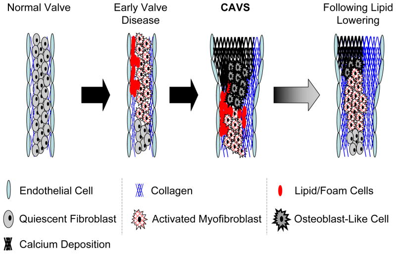
Figure 6.
Progression and “regression” of CAVS in “Reversa” mice12, 13. Early stages of CAVS in mice involve myofibroblast activation and lipid insudation/foam cell formation, and are followed by the appearance of osteoblast-like cells, valvular calcification, and substantial increases in valvular fibrosis. Following reduction of blood lipids (“regression” in right panel), there are substantial reductions in valvular lipid content and calcium content, but valvular fibrosis remains increased. Despite reduction of valvular lipid and calcium content, aortic valve function does not improve with substantial lipid lowering.

