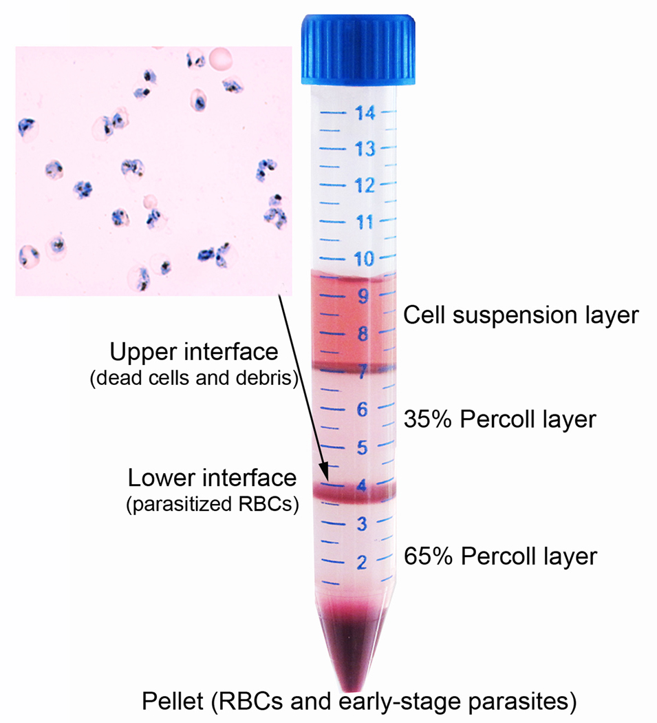
Figure 1.
Purification of parasitized RBCs on a step Percoll gradient consisting of an upper 35% and a lower 65% Percoll layer. After centrifugation, the upper interface contains mostly cell debris, whereas the lower interface contains enriched parasitized RBCs. The bottom pellet contains mostly unparasitized RBCs and ring-stage parasites. Inset shows an image from Giemsa-stained thin smear from the lower interface, which contains mostly late-stage trophozoites.

