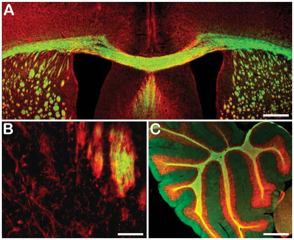
Figure 5.
Ex vivo BDB staining of myelin sheaths in the corpus callosum (green in A) and cerebellum (green in C) colocalized with MBP staining (red) in the same sections. However, ex vivo staining of oligodendrocyte cell soma present in caudate putamen with BDB showed lack of staining in the cell bodies (B), whereas immunostaining for MBP was positive, indicating that BDB preferentially stains myelinated fibers. Bars: A,C = 500 μm; B = 50 μm.

