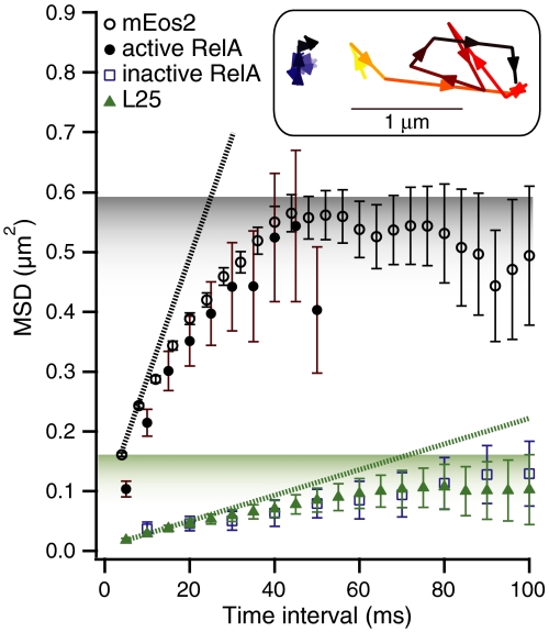
Fig. P1.
Diffusion comparisons. Mean square displacements (MSDs) for mEos2 (in black), ribosomal protein L25 (in green), active RelA (in brown, with 2.5 mM of L-Serine Hydroxamate), and for inactive RelA diffusion (in blue, in exponentially growing cells) with error bars representing standard errors of the means. Inactive RelA diffusion is indistinguishable from the diffusion of Dendra2-labeled ribosomes. When cells are starved, the diffusion of RelA changes dramatically and is similar to that of mEos2. (Inset) When the temperature is held constant, only slowly diffusing RelA trajectories are observed (left trajectory, recorded with a frame time of 10 ms). During the temperature jump from 21 °C to 37 °C, one transiently observes fast diffusion trajectories of RelA (right trajectory, with a frame time of 10 ms).

