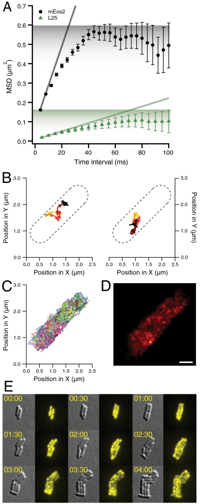
Fig. 3.
Single-molecule ribosome tracking and ensemble time-lapse imaging in individual living cells. (A) Mean square displacements (MSDs) in the sample (x-y) plane for mEos2 (in black, from 3,766 trajectories) and ribosome (in green, from 537 trajectories) tracking for different time intervals with error bars representing standard errors of the means. For ribosome tracking, we labeled the ribosomal protein L25 with the photoconvertible GFP variant Dendra2 (25) (see Materials and Methods). The dotted lines are calculated from the slope of the first two time points (corresponding to Dapp of 5.1 and 0.5 μm2 s-1, respectively). As estimated from the initial slopes of the MSD curves, mEos2 has a 10-fold higher apparent diffusion coefficient than Dendra2-labeled ribosomes. The shading represents the different plateaus for mEos2 and L25 MSDs. (B) Two experimentally obtained single-molecule ribosome trajectories with a frame time of 50 ms. The individual ribosome trajectories are recorded for 0.65 and 1.15 s, respectively. The ribosome is tagged via an N-terminally Dendra2 labeled L25 ribosomal protein. (C) Overlay of all 224 single-molecule ribosome trajectories in one E. coli cell with a frame time of 50 ms. (D) Overlay of 1,000 positions of single-molecule ribosome trajectories in one E. coli cell with a frame time of 50 ms. Each position is represented by a Gaussian with a standard deviation equal to the localization error. The mean localization error is 43 nm and the scale bar represents 500 nm. (E) Time-lapse image acquisitions (differential interference contrast and fluorescence imaging) of ribosome distributions in dividing E. coli cells. The fluorescence of Dendra2 from chromosomally labeled C-terminal ribosomal protein S2 is activated at time zero. Subsequent time-lapse imaging followed the initial distribution of the photoconverted ribosomes as they are passed between the cells upon repeated cell division. The cellular distribution of this photoconverted Dendra2 is recorded every five minutes. We present nine snapshots over a period of four hours. Experiments with ribosomal protein L19 and L31-labeled strains resulted in similar data.

